
Cellular senescence is the permenant cell cycle arrest to response to various cell stress. The accumulation of senescence is considered to contribute to the process of aging disease, including chronic lung aging disease. NIH launches program to map a rare type of non-dividing cells implicated in human health and disease. This is a consortium collaboration project and funded by NIH, leaded by Prof. Toren Finkel, Prof. Melanie Koenigshoff, Prof Ana Mora and Prof. Irfan Lucia Rahman from three states of USA. The consortium will identify biomarkers of senescent cells in human heart and lung and then construct high-resolution, detailed maps of cellular senescence across the lifespan and physiological states. Our analysis will involve in situ mapping using established targeted assays, as well as high-content unbiased approaches involving single cell RNA/ATAC sequencing, proteomics, and spatial transcriptomics. In addition, as part of our mapping endeavor, the TriState SenNet TMC will analyze the relationship between the initiating trigger for senescence and the subsequent biology and phenotype of the senescent cell.
This was the doctoral research for doctoral degree.
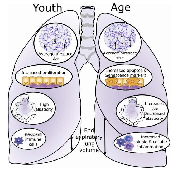
As a doctoral candidate, I have gotten a hybrid training on computational skills and biological skills. My research contributions focused on epithelial cell reprogramming in lung aging and lung fibrosis. More specifically, I phenotyped the lung epithelial cells and identified cellular senescence as a common type of reprogramming in both alveolar and airway regions during lung aging and lung fibrosis. I compared the cell population in young and old mice and demonstrated declined population of epithelial cells (EpCAM+) in old mice. Further analysis demonstrated increased activity of β-catenin and senescence-associated β-galactosidase (SA-β-GAL) in primary AT2 cells in aging mice. Similarly, I compared epithelial cell subpopulation in lung tissue from IPF patients and health donors and revealed an enlarged population of airway epithelial cells and sharply declined population of AT2 cells in IPF patients. I further identified G-protein coupled receptor 87 (GPR87) as a novel marker of airway basal cells within honeycombs cysts in IPF. Then I investigated the role of developmental pathway within both alveolar and airway regions and demonstrated the activation of developmental signaling pathway contributing to epithelial cell senescence. My work highlighted the significance and connection of developmental signaling pathway and cellular senescence during the development of IPF. Furthermore, I generated two datasets of single cell RNA-sequencing, with one from lung tissue of young healthy donors and old healthy donors, the other one from lung tissue of health donors and IPF patients. Both datasets were enriched with EpCAM+ cells isolated from parenchymal lung tissue, providing high-resolution of epithelial cell atlas to the research community of lung aging and lung fibrosis.
This was the graduate research for my Master's degree.
In this project, I developed a method, which was proved to be of feasibility and reliability, to quantitatively high-throughput dissect the cellular microenvironment in microfluidic culture systems. This method was based on LC-MS/MS and microfluidics technology. By miniaturization of the culture system within a microfluidic chip, it could have a different balance between endogenous factors (secreted by the cells) and exogenous factors (provided by the culture medium).
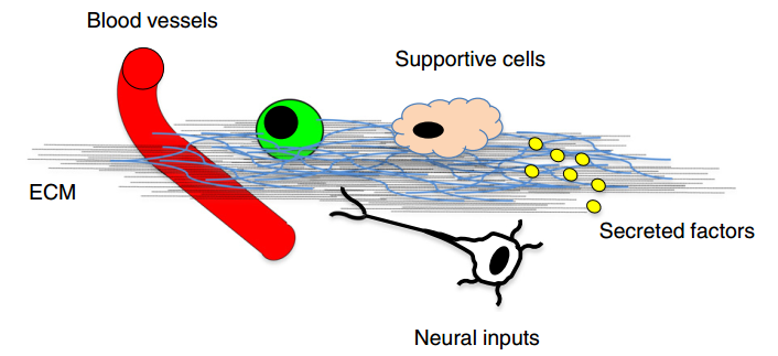 Using this method, I explored the cellular microenvironment of human induced pluripotent stem cells (hiPSCs) from different aspects, including the soluble secreted proteins and composition of extracellular matrix (ECM), identifying about 600 proteins with 66.2% being extracellular proteins. I further dissecting the cellular microenvironment during cell reprogramming. The structure of the ECM showed different remodeling during cell reprogramming within microfluidics and normal well system. Considering the huge disparity of reprogramming efficiency between these two systems, we concluded that ECM is likely affecting cell reprogramming efficiency, while cell reprogramming process under different environment could also remodel the ECM.